What is a Cholesteatoma?
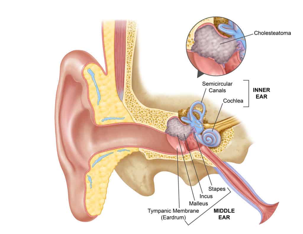
A cholesteatoma is an abnormal skin growth in the middle ear behind the eardrum. Repeated infections can allow skin to build up in the middle ear. The growth, however, is not cancerous.
Cholesteatomas often develop as cysts or pouches that shed layers of old skin inside the middle ear. Over time, the cholesteatoma can grow and damage the surrounding delicate bones of the middle ear. This can lead to hearing loss that surgery can often improve.
Ongoing growth into the vital structure of the ear bone can lead to a permanent loss of hearing, balance and facial nerve function, so detection and treatment is important.
What Causes a Cholesteatoma?
A cholesteatoma is usually caused by infection in the middle ear and poor eustachian tube function. The eustachian tube sends air from the back of the nose into the middle ear to equalize ear pressure or “clear the ears”.
When the eustachian tubes work poorly, sometimes due to allergy, a cold, or sinusitis, the air in the middle ear is absorbed, creating a partial vacuum in the ear. The pressure sucks in a pouch or sac by stretching the eardrum — especially areas weakened by previous infections. This can develop into a sac and become a cholesteatoma.
A rare congenital form of cholesteatoma (one present at birth) can occur in the middle ear and elsewhere, such as in the nearby skull bones. However, the type of cholesteatoma associated with ear infections is most common.
How is a Cholesteatoma Treated?
An examination by an otolaryngologist (a ear, nose and throat doctor) can confirm a cholesteatoma. Initial treatment may consist of carefully cleaning the ear, antibiotics, and ear drops. Therapy can also stop drainage in the ear by controlling the infection. At this stage, the cholesteatoma’s must be evaluated.
Most cholesteatomas usually requires surgical treatment to protect the patient from serious complications. Hearing and balance tests and CT scans may be necessary to determine the hearing level and the damage caused.
In most cases, surgery is performed under general anaesthesia. The main purpose is to remove the cholesteatoma and eliminate the infection so the ear can remain dry. A second surgery is sometimes needed to ensure the cholesteatoma is gone and to attempt reconstruction of any damaged middle ear bones to improve hearing.
In cases of severe ear damage, reconstruction may not be possible, or it may require another operation six to 12 months later. The operation will allow the surgeon to inspect the middle ear space and mastoid for residual cholesteatoma.
Surgery is performed as an inpatient in hospital. An overnight stay is usually necessary and seven to 10 days off work generally required. After surgery, follow-up office visits will evaluate results and check for recurrence. Some patients will need lifelong, periodic ear examinations.
Symptoms and Dangers
Cholesteatoma symptoms include:
- A feeling of fullness or pressure in the ear, along with hearing loss (caused by the cholesteatoma pouch or sac growing);
- The ear draining fluid with a foul odour; or
- Dizziness or muscle weakness on one side of the face (the side of the infected ear).
Any or all of these symptoms are good reasons to seek medical evaluation.
Cholesteatoma is a serious but treatable ear condition which can be diagnosed only by medical examination. Bone erosion can cause the infection to spread into the surrounding areas including the inner ear and brain, and as such, choleasteatoma should be treated as soon as possible.
Cholesteatoma Publications
Professor Marcus Atlas is a world-leading expert in cholesteatomas and has published many peer reviewed international articles and books on the surgery of this disorder.
Reference
Cholesteatoma Web 3-23-11, American Academy of Otolaryngology-Head and Neck Surgery Patient Information Leaflet
63. Singh V, Atlas MD. Obliteration of the persistently discharging mastoid cavity using the middle temporal artery flap. Otolaryngol Head Neck Surg. 2007 Sep;137(3):433-8
BOOK
Atlas M, Eisenberg R, (eds). A Guide to Temporal Bone Dissection (2nd Ed.). Perth: Lions Ear & Hearing Institute, 2004.
DVD
Atlas M. Ear and Temporal Bone Surgery – A DVD Guide, Vol 1: Soft Tissue Approach. Perth: Lions Ear and Hearing Institute, 2005.
Atlas M. Ear and Temporal Bone Surgery – A DVD Guide, Vol 2: Middle Ear and Mastoid Surgery. Perth: Lions Ear and Hearing Institute, 2005.
Atlas M. Ear and Temporal Bone Surgery – A DVD Guide, Vol 3: Neurotology. Perth: Lions Ear and Hearing Institute, 2005.